
Overview
Osteoarthritis (OA) is the most common form of arthritis (joint disease) and one of the leading causes of pain and disability worldwide. The knee is the most commonly affected joint with symptomatic patients reporting pain accompanied by varying degrees of functional limitation and reduced quality of life. Knee OA can be diagnosed using the patient’s age, symptoms and a physical examination, with or without the addition of X-rays. Exercise, education and weight loss improve symptoms and function in most patients with knee OA and should be trialled before considering surgery. Surgery is an effective treatment option but carries greater potential risks and may be deemed unsuccessful by a small proportion of patients.
Disease process
Knee OA is often described as ‘wear and tear’, implying that the joint has ‘worn out’ with use like the sole of a shoe. Symptoms and physical function are assumed to be directly related to structural changes within the joint, all of which would logically deteriorate with further use. On the contrary, appropriately loading the knee stimulates tissue regeneration and is essential for joint health. Structural changes do not always correspond with symptoms and there is variability in the progression of signs, symptoms and function between individuals with knee OA.
Structurally, an abnormality in any of the knee tissues (e.g. cartilage, ligament, meniscus) can increase stress within the joint, initiating tissue damage and repair. The repair process is slow but efficient and can often compensate for the initial damage. Biomechanical, biochemical and/or genetic factors can influence joint stress and inhibit repair, which ultimately results in the breakdown of cartilage and bone within the joint. From a symptom perspective, some patients with knee OA will progressively worsen while others will demonstrate either non-progressive or improving trajectories. For those with improving symptoms, there may be a corresponding increase in function, while for others, function may deteriorate significantly as certain activities are avoided due to actual or anticipated pain.
Knee OA therefore involves multiple joint structures with symptoms and function likely influenced by a combination of structural, psychological and behavioural factors.
Risk factors
There are numerous factors that are associated with knee OA. Risk factors such as age, female gender, previous knee injury and family history of OA cannot be modified. Modifiable factors include:
Weight: being overweight or obese is an important factor in the development and progression of knee OA.
Muscle weakness: quadriceps weakness is a risk factor for developing knee OA.
Future knee injury: injury prevention programmes have been shown to be effective in reducing knee injuries that are likely to result in knee OA (e.g. anterior cruciate ligament injury).
Presentation
Knee OA patients can present with variable signs and symptoms; the severity of symptoms can fluctuate within the same individual. While some patients will have symptoms that worsen over time, others can have mild, non-progressive or improving symptom trajectories.
In early presentations, pain is usage-related, relieved with rest but often feels worse at the end of the day. If present, knee joint stiffness is experienced after periods of inactivity, particularly after getting out of bed (early morning stiffness), but resolves within 30 minutes of movement. There may be modest swelling within the joint (effusion), a grating sensation (crepitus) whilst actively moving the knee (e.g. squatting) or a sensation of the knee ‘giving way’.
In advanced presentations, pain can be more pronounced at night or persist at rest. The person may no longer be able to fully bend or straighten their knee (restricted range of movement), bones may be enlarged and the legs can appear ‘bow-legged’ or ‘knock-kneed’.
The associated signs and symptoms of knee OA can result in functional limitations (e.g. reduced walking distance) and reduced quality of life.
Diagnosis
Knee OA can be diagnosed using the individual’s age, symptoms and a physical examination, as described below. For details on the diagnostic accuracy of clinical signs and symptoms for knee OA, please visit the statistics section.
NICE guidelines:
The National Institute for Health and Care Excellence (NICE) Guidelines for Osteoarthritis (2014) state that a diagnosis of OA can be made without investigations if a person:
- is aged 45 years or over AND
- has activity related joint pain AND
- has either no morning joint-related stiffness or morning stiffness that lasts no longer than 30 minutes.
EULAR guidelines:
The European League Against Rheumatism (EULAR) guidelines state that a confident diagnosis of knee OA can be made without X-ray if an adult >40 years old has (1) usage-related pain, (2) short-lived morning stiffness, (3) functional limitation AND one or more of the following examination findings: (4) crepitus, (5) restricted movement or (6) bony enlargement. This applies even if X-rays have been taken and appear normal.
Patients ≥45 years old presenting with all 6 signs and symptoms have a 99% probability of having knee OA.
ACR criteria:
The American College of Rheumatology (ACR) uses clinical findings, with or without X-ray and laboratory findings. Unlike the NICE and EULAR guidelines, the ACR criteria for the classification of OA also accounts for patients under the age of 40. Using clinical findings alone, a patient with symptomatic knee OA should have knee pain AND at least three of the following 6 criteria:
- age >50 years
- morning stiffness <30 minutes
- crepitus on active movements
- tenderness of the bony margins of the joint
- bony enlargement
- no palpable warmth.

Classification
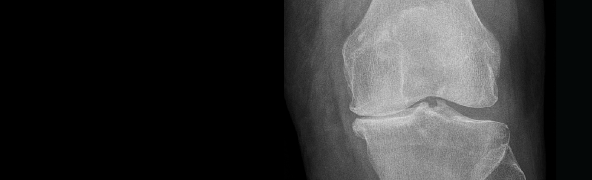
Numerous knee OA grading scales have been proposed based on X-ray findings. The Kellgren & Lawrence classification system is the most commonly used scale but has been interpreted and described in various ways. To grade tibiofemoral OA, plain anterior-posterior (AP) weight bearing X-rays are used (images 1-5). Rosenberg or Schuss views are posterior-anterior (PA), weight bearing X-rays performed with the knee bent to 45° and 30° respectively; both views are more sensitive than plain weight-bearing X-rays for detecting joint space narrowing. Side (lateral) and skyline views are required to grade patellofemoral joint OA (images 6-9).
Knee OA is graded by the presence of osteophytes, joint space narrowing, hardening of bone beneath the cartilage (subchondral bone sclerosis) or deformity of the bone ends.
Grade:
0: No joint space narrowing or reactive changes (image 1)
1: Doubtful joint space narrowing, possible osteophytic lipping (image 2)
2: Definite osteophytes (white arrows), possible joint space narrowing (image 3)
3: Moderate osteophytes, definite joint space narrowing, some sclerosis, possible bone end deformity (image 4).
4: Large osteophytes, marked joint space narrowing, severe sclerosis, definite bone ends deformity (image 5).


Images 1-5: Grade 0-4 Kellgren & Lawrence classification of knee OA


Images 6-7. Lateral and skyline views showing patellar osteophytes (white arrows) but well-preserved patellofemoral joint space.
Images 8-9. Lateral and skyline views showing patellar (white arrows) femoral osteophytes with marked patellofemoral joint space narrowing.
It is important to note that X-ray findings and symptoms do not always correspond well. Patients with definite knee OA changes on X-ray may have no symptoms, while patients with severe symptoms may have negligible findings on X-ray. In the presence of X-ray evidence of knee OA, a diagnosis of knee OA can only be made if the patient describes knee pain. 99.5% of X-rays do not reveal anything to suggest the core treatments for knee OA are inappropriate.
Treatment
Education, exercise and weight management are considered the “core” treatments for knee OA and should be offered to all patients, irrespective of symptom severity, age, disability or co-existing medical conditions. These interventions have been shown to postpone knee surgery in 3 out of 4 patients for at least one year, 2 out of 3 patients for at least two years, and provide clinically-relevant improvements for most people, without the associated risks of total knee replacement.
Education:
Pain is a complex topic that is influenced by an individual’s beliefs, therefore patients should be offered accurate information regarding the disease process and management of knee OA, addressing any misconceptions they may have. Pain is also influenced by other factors including mood, sleep and coping strategies. Self-management strategies should be developed with the individual, ensuring positive lifestyle changes are implemented alongside the core treatments and that they are maintained in the long-term.
Exercise:
Exercise is at least as effective as non-steroidal anti-inflammatory drugs (NSAIDs) and 2-3 times more effective than paracetamol for reducing pain in knee OA. Optimal exercise programs for knee OA should have one aim; focus on improving aerobic capacity, quadriceps muscle strength, or lower extremity performance. For best results, the program should:
- be supervised
- be performed 2-3 times per week
- consist of at least 12 sessions.
If both aerobic endurance and strength are indicated, exercises should be performed on separate days. Any benefits gained from exercise are likely to remain if the exercises are continued, but disappear when they are stopped, therefore adherence to a long-term exercise programme is important.
Exercise prescription should be individualised. It is important to choose a modality that an individual can easily access, is likely to persist with and does not cause an excessive flare up of symptoms; small flare ups of pain are acceptable, provided they settle within 24 hours. If weight bearing exercises are not tolerated, lower weight bearing alternatives should be trialled (e.g. aquatic exercise or bike).
It is unclear which patients will benefit from specific forms of exercise but emerging evidence suggests quadriceps strengthening may be more effective in obese patients, while lower extremity performance may be more effective in those with medial knee OA and a varus thrust.
Aerobic exercise:
Aerobic exercise aims to increase heart rate and oxygen uptake. This can be achieved by performing different activities including swimming/aquatic exercises, cycling or walking for at least 10 minutes.
Strength exercises:
For clinically significant improvements in pain and disability, individuals should aim for at least 30% improvement in strength. This can be achieved by using different resistance exercises including knee extension, leg press, or squatting.
The American College of Sports Medicine (ACSM) resistance training guidelines for muscular strength are as follows:
- Load: 60-70% 1RM for novice to intermediate; 80-100% for advanced
- Volume: 1-3 sets of 8-12 repetitions for novice to intermediate; 2-6 sets of 1-8 repetitions for advanced
- Rest period: 2-3 min for higher intense exercises that use heavier loads; 1-2 minutes between the lower intense exercises with light loads
*1RM = one repetition maximum.
Resistance exercises will need to be performed for at least 2-3 months to achieve the desired improvements in strength.
Functional/lower limb performance:
Neuromuscular exercise (NEMEX) training aims to improve the quality and efficiency of movement, which may be impaired in patients with knee OA, whilst also incorporating components of strengthening. The principle of NEMEX training is to maintain neutral alignment of the leg during progressively more challenging functional tasks. These exercises are demonstrated on the GLA:D international YouTube page, and can be accessed by clicking here.
Weight management:
Being overweight or obese is a risk factor for the development and progression of knee OA. Patients are classed as overweight if their body mass index (BMI) is between 25-30kg/m2, or obese if their BMI is ≥ 30kg/m2.
In overweight or obese patients, losing weight has been shown to improve pain, function and the appearance of joint cartilage. For every unit of weight lost/gained, there is a four-fold decrease/increase in knee joint load per step; accumulated over thousands of steps per day, this is likely to be clinically meaningful for individuals with knee OA. A 5% reduction in body weight can improve function, with less predictable effects on pain relief, while a 10% loss in weight can lead to substantial improvements in pain. There is a dose-response relationship between weight and symptoms; the more weight is lost, the more symptoms improve.
Weight loss is most likely to be achieved by combining exercise and diet. Individuals with knee OA, that have a BMI less than 25kg/m2, should ensure they do not put on excessive amounts of weight.
Adjuncts
If required, adjunct treatments should be used alongside the core treatments, not in place of them. The Osteoarthritis Research Society International (OARSI) describe the following interventions as ‘appropriate’ for knee OA.
Knee braces: in cases of medial compartment knee OA with varus (bow-legged) knee alignment, low-quality evidence shows benefits of a medial unloader brace for pain, stiffness, function and quality of life (image 10-12).


Images 10-12: unloader braces for medial knee OA and varus knee alignment.
Corticosteroid injections (CSI): CSI into the knee joint may produce a moderate improvement in pain and small improvement in physical function. Any beneficial effects gained from injection are likely to decrease over time and are unlikely to remain after six months of being administered. Performing an injection prior to exercise for patients with painful knee OA does not improve outcomes when compared with exercise alone.
Injection therapy is also associated with specific adverse events, the most serious being an allergic reaction to a drug (anaphylaxis) or an infection within the joint (septic arthritis). CSI repeated every 3 months for 2 years has recently been shown to reduce cartilage volume, with no significant difference in knee pain, when compared with saline injections. Repeated use of CSI every three months is therefore not supported based on this information.
A cane/walking stick and topical non-steroidal anti-inflammatories (NSAIDs) are appropriate for individuals with knee OA only. Oral NSAIDs and capsaicin are appropriate for patients without relevant co-morbidities, but are associated with known side-effects. Paracetamol (acetaminophen), has historically been recommended for symptom relief but recent, high-quality evidence suggests this drug is clinically ineffective and should not be recommended for knee OA, irrespective of the dose.
Surgery
Arthroscopic surgery is not superior to exercise therapy for knee OA and is associated with rare, but potentially serious complications (0.5% blood clot, 0.2% infection). Knee arthroscopy is therefore not recommended for knee OA patients, even in the presence of mechanical symptoms (catching/locking).
High tibial osteotomy (HTO) (image 13), distal femoral osteotomy and uni-compartmental knee replacement (UKR) (image 14-15) are surgical options for knee OA that is confined to one compartment of the knee. Although neither procedure has been proven to be superior to the other for medial knee OA, valgus HTO provides better physical activity for younger patients whereas UKA is more suitable for older patients due to shorter rehabilitation time and faster functional recovery. The potential unloading effect of a valgus HTO may be predicted by trialling a medial unloader brace (images 10-12) before surgery; this is referred to as the ‘brace-test’.
Total Knee Replacement (TKR) (image 16), followed by core treatments, is considered an effective treatment for end-stage knee OA. Compared with core treatments alone, TKR and core treatments provide greater improvements in pain relief, function and quality of life. However, TKR is associated with more serious adverse events, including blood clots, infection, knee stiffness and fracture.


Images 13-16: X-ray images of valgus high tibial osteotomy, unicompartmental knee replacement, patellofemoral joint replacement and total knee replacement.
Acknowledgements
Written by: Richard Norris, The Knee Resource
Reviewed by: Søren Thorgaard Skou PT, PHD
Professor
Research Unit for Musculoskeletal Function and Physiotherapy
University of Southern Denmark
Head of Research
Department of Physiotherapy and Occupational Therapy
Næstved-Slagelse-Ringsted Hospitals
Twitter: @STSkou

References
Altman R, Asch E, Bloch D, Bole G, Borenstein D, Brandt K, et al. Development of criteria for the classification and reporting of osteoarthritis. Classification of osteoarthritis of the knee. Diagnostic and Therapeutic Criteria Committee of the American Rheumatism Association. Arthritis Rheum. 1986;29(8):1039-49.